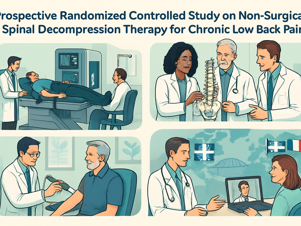
A prospective randomized controlled study of non surgical spinal decompression therapy for the treatment of chronic low back pain
December 16, 2011
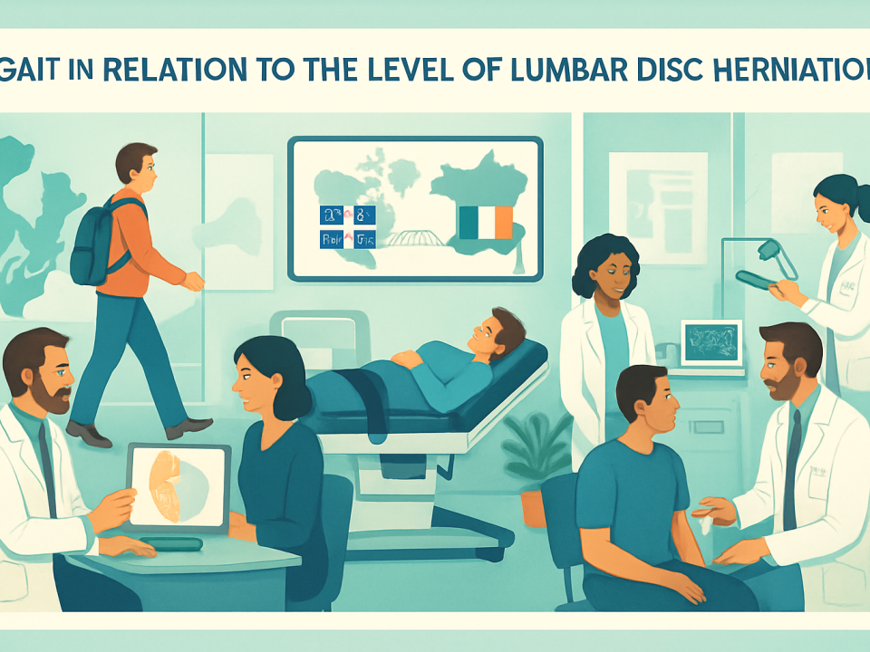
Gait in relation to the level of lumbar disc herniation
December 19, 2011
The prevalence of lumbar disc herniation remains a significant concern in musculoskeletal and neurological health worldwide. Emerging as a prevalent source of low back pain, it poses diagnostic challenges and demands nuanced clinical and radiographic evaluation to optimize patient outcomes. Understanding the specific signs, both clinical and imaging-based, which indicate not only the presence but also the degree of lumbar disc herniation is crucial for informed care pathways. From subtle changes in lumbar motion to advanced imaging techniques such as MRI and CT scans, a comprehensive approach helps pinpoint hernias’ impact on nerve roots and spinal stability. Experts like Dr. Sylvain Desforges, with over 30 years of experience, emphasize the integration of evidence-based diagnostics with patient-centered evaluation to tailor non-surgical and surgical interventions. This article unpacks the latest consensus and clinical insights about optimum signs of lumbar disc herniation, guiding better diagnosis and personalized treatment strategies, while highlighting innovations shaping modern spine care.
Comprehensive Epidemiology and Risk Profile of Lumbar Disc Herniation
Lumbar disc herniation affects approximately 1–3% of the population symptomatically during their lifetime, with an overall lifetime risk estimated at up to 30%. According to the World Federation of Neurosurgical Societies (WFNS), most cases resolve spontaneously, highlighting the body’s capacity for natural recovery. Understanding who is at risk involves examining genetic predispositions, occupational hazards, and lifestyle factors.
Men between 30 and 50 years old constitute the demographic most frequently affected, with herniations predominantly at the L4-L5 and L5-S1 levels. Occupations involving strenuous physical activity or prolonged heavy lifting increase risk, including professions such as surgeons, professional drivers, and athletes. Smoking has also been demonstrated to promote disc degeneration and herniation significantly.
In a landmark 33-year prospective study from Copenhagen, strenuous work-related activity was the strongest predictor for hospitalization due to lumbar disc herniation, underscoring the environmental component. However, genetic influence is substantial and may outweigh occupational factors, as twin studies have shown that inherited traits govern susceptibility to disc degeneration and subsequent herniation.
Body composition specifics, such as increased visceral fat and abdominal circumference, have emerged as contributory risk factors beyond simple body weight metrics. This complexity explains the diverse presentation and progression among patients.
Risk Factors Summary:
- Genetic predisposition influencing disc degeneration.
- Male gender, especially aged 30-50 years.
- Physical activities involving heavy lifting or repetitive strain.
- Cigarette smoking accelerating disc pathology.
- Visceral obesity contributing to mechanical and metabolic stress on discs.
These risk factors guide the clinician’s index of suspicion, influencing the urgency and type of diagnostic assessments such as clinical examinations and imaging studies. For further reading on epidemiology and guidelines, the full WFNS consensus and detailed epidemiological data can be accessed at NCBI Bookshelf and related resources like The Radiology Assistant’s lumbar disc herniation overview.

Gait in relation to the level of lumbar disc herniation
The complex relationship between lumbar disc herniation and gait abnormalities presents an important dimension in understanding spinal health and functional mobility. Lumbar disc herniation, a prevalent orthopedic condition, often manifests in altered walking patterns due to neurological deficits and musculoskeletal…
Clinical Manifestations and Pain Evaluation in Lumbar Herniated Discs
Patients with lumbar disc herniation commonly present with low back pain radiating into one or both legs, a manifestation of radiculopathy caused by nerve root irritation or compression. However, clinical presentation is diverse, and history taking forms the cornerstone of evaluation.
Key aspects of pain assessment include intensity, onset, localization, and associated neurological symptoms such as numbness, weakness, or muscle spasms. Validated measurement tools such as the Numerical Pain Rating Scale (NPRS) and the Oswestry Disability Index (ODI) help quantify patient suffering and functional impairment, facilitating treatment decisions.
Discogenic pain from annular tears or nucleus pulposus displacement commonly exhibits centralization signs, differentiating it from facetogenic or sacroiliac joint pain. Sometimes, provocation discography is used to identify discogenic pain, though its role is controversial due to risk of accelerated degeneration and false positives.
Physical examination incorporates essential tests such as:
- Manual muscle testing using the Medical Research Council (MRC) scale.
- Sensory examination of dermatomal distribution.
- Evaluation of reflexes and sphincter function.
- Straight leg raise (Lasegue sign) and crossed Lasegue sign to elicit radicular symptoms.
Notably, a positive crossed Lasegue sign strongly suggests nerve root irritation and is proportionate to the degree of herniation. However, isolated neurological signs may not reliably predict herniation level or grade. Hence, these tests must be integrated with patient history and imaging findings for accurate diagnosis.
For an in-depth understanding of clinical signs and evaluation techniques, refer to resources such as Which Orthopedic Examinations Reveal a Herniated Lumbar Disc and Dr. Desforges’ insights on herniated disc symptoms.

Chronic low back pain affects millions globally, prompting the development of diverse treatment strategies. Among these, non-surgical spinal decompression therapy has emerged as a prominent conservative option, acclaimed for its potential to alleviate pain without invasive procedures. This treatment aligns…
Optimizing Radiologic Diagnosis: MRI and CT Scan in Lumbar Disc Herniation
Magnetic resonance imaging (MRI) is recognized universally as the gold standard for diagnosing lumbar disc herniation due to its excellent soft tissue contrast and detailed assessment of intervertebral discs, nerve roots, and surrounding structures. MRI enables visualization of the nucleus pulposus displacement, annular tears, and the presence of T2 hyperintensity, indicative of inflammation or edema.
Typical MRI features suggestive of herniation include:
- Disc bulge extending beyond normal margins.
- Fragmentation or extrusion of herniated disc fragments compressing nerve roots.
- Annular tears visible as high-intensity zones.
- Contrast enhancement signaling inflammation or nerve root irritation.
- Sagittal imaging for comprehensive evaluation of disc height and spinal alignment.
Computed tomography (CT) and CT myelography serve as supplemental imaging techniques when MRI is contraindicated or inconclusive. CT provides excellent bone detail aiding detection of foraminal osteophytes or calcifications and can elucidate nerve root compression visually, especially valuable for grading radiculopathy severity.
Diagnostic accuracy varies depending on the imaging technique and grade of herniation. Multidetector CT has demonstrated sensitivity and specificity rivaling or exceeding older MRI technology, though advances in high-field MRI have narrowed this gap. Importantly, evaluations should consider common asymptomatic findings such as disc bulges, which appear on imaging in a significant portion of adults over 50 without painful symptoms, underscoring the importance of correlating clinical and radiologic data.
| Imaging Modality | Sensitivity Range | Specificity Range | Optimal Usage |
|---|---|---|---|
| MRI | 75% – 97% | 77% – 99% | First line for soft tissue, nerve root visualization |
| CT Scan | 77% – 98% | 73% – 96% | Bone detail, contraindicated MRI cases |
| CT Myelography | 75% – 98% | 76% – 96% | When MRI inconclusive or not possible |
References for detailed neuroradiological characterization are available at Classification and Diagnostic Imaging of Herniated Discs and Dr. Desforges’ herniated disc diagnosis guide.

CLINICAL STUDIES ON THE EFFECTIVENESS OF SPINAL DECOMPRESSION
Spinal decompression therapy stands at the forefront of non-invasive treatments for chronic low back pain, a condition that persists as a major health challenge worldwide. Affecting up to 85% of individuals during their lives, low back pain not only diminishes…
Correlation Between Clinical Signs and Imaging Findings For Accurate Herniation Grading
Correctly grading the degree of lumbar disc herniation is pivotal in choosing appropriate management paths. Studies indicate that certain clinical signs correlate strongly with the severity of herniation as detected by imaging.
The degree of herniation can be described as:
- Protrusion (bulge): Disc material bulging beyond the disc margin but contained by the annulus fibrosus.
- Extrusion: Herniated material breaches the annulus but remains attached to the disc.
- Sequestration: Free fragment of the herniated disc migrates and separates completely from the parent disc.
The range of lumbar sagittal motion diminishes progressively with increasing severity of herniation; barely any motion is present in protruded hernias with root involvement, and further restrictions appear with extrusion and sequestration. The crossed Lasegue sign’s presence proportionally reflects the grade of herniation; when combined with lumbar range-of-motion findings, clinical assessments can predict uncontained (sequestrated) hernias in up to 74% of cases and contained hernias (protruded and extruded) in 68%.
Neurologic signs alone, such as reduced Achilles reflex, may not reliably localize the herniation level but help distinguish radicular pain from other etiologies. Advanced clinical evaluation combined with detailed MRI or CT scans enables a comprehensive assessment of nerve root compression severity, which is essential for personalized treatment strategies.
For detailed clinical evaluation techniques linked with radiographic findings, readers may explore Dynamic Chiropractic’s review on optimum signs and Dr. Desforges’ publications at muscle function abnormalities in lumbar herniation.
Nonsurgical Spinal Decompression To Treat Chronic Low Back Pain
Chronic low back pain remains one of the most pervasive health challenges worldwide, affecting millions and severely impairing quality of life. As individuals seek alternatives to invasive surgeries, nonsurgical spinal decompression therapy emerges as a pioneering approach offering hope for…
Innovative Non-Surgical Treatments Guided by Diagnostic Clarity
Non-surgical spinal decompression therapies have gained traction as effective alternatives for carefully selected patients with lumbar disc herniation. Accurate diagnosis through clinical and imaging markers ensures the ideal candidates are identified, optimizing treatment success rates while avoiding unnecessary invasive interventions.
Dr. Sylvain Desforges advocates integrating advanced diagnostics with state-of-the-art therapies such as:
- Mechanical spinal decompression targeting herniated discs to reduce nucleus pulposus pressure.
- Laser therapy to promote tissue repair and reduce inflammation linked to annular tears.
- Shockwave treatment supporting healing of degenerative changes.
- Dynamic spinal implants preserving mobility post-intervention.
The rationale behind these treatments involves decompressing nerve roots affected by herniated disc fragments and promoting natural resorption and healing of displaced nucleus pulposus tissue. Positive outcomes from conservative therapy typically manifest within 6 to 12 weeks, aligning with guidelines recommending radiologic re-evaluation for persistent symptoms.
Evidence-based clinical pathways emphasize thorough initial conservative management before considering surgery, enhancing patient quality of life sustainably. More information on these therapeutic options and eligibility can be found on Dr. Desforges’ comprehensive decompression therapy guide and the latest innovations showcased at TAGMED Clinic’s prosthetic approaches in Montreal.

Spinal decompression outcome of clinical study
In recent years, spinal decompression therapy has emerged as a promising non-invasive treatment for patients suffering from herniated discs and other degenerative spinal conditions. The efficacy of this approach is under continual scrutiny through clinical studies evaluating both immediate and…
International Collaboration Enhancing Patient Care Across Borders
The management of lumbar disc herniation increasingly benefits from transatlantic cooperation between Canadian and French healthcare teams, exemplified by collaborations involving Dr. Sylvain Desforges and well-known medical platforms like SOS Tourisme Médical. This model facilitates access to advanced diagnostics, specialized interventions, and comprehensive follow-up care.
Patients benefit from:
- Streamlined referral pathways between Montreal and France.
- Access to surgical techniques and dynamic implants currently unavailable or limited in Canada.
- Coordinated care plans ensuring timely consultations and personalized treatment trajectories.
- Sharing of research, training, and technological advancements between multidisciplinary teams.
This approach exemplifies a patient-centered, evidence-driven philosophy promoting optimal lumbar disc herniation outcomes while respecting local medical regulations and ethical standards. Cross-national care coordination improves quality and mitigates long surgical wait times often experienced in public health systems.
Prospective patients seeking such integrated care are encouraged to initiate contact through trusted platforms detailed at SOS Tourisme Médical contact page and explore Dr. Desforges’ role in fostering this transatlantic exchange.

Candiac Back Pain Relief for Herniated Disc Sufferers
Back pain stemming from herniated discs remains a challenging and widespread issue impacting many individuals in Candiac and the surrounding Quebec regions. This condition often leads to persistent discomfort, restricted mobility, and diminished quality of life for sufferers. In the…
Patient Education: Empowerment Through Understanding Lumbar Disc Herniation
Clear communication about the nature, implications, and management options of lumbar disc herniation is vital for patient empowerment. Simplifying complex terminology such as the difference between a herniated disc fragment and an annular tear, or explaining why MRI findings may not always align with symptoms avoids confusion and encourages active participation in care planning.
Education efforts focus on:
- Clarifying the significance and limitations of imaging results like T2 hyperintensity and contrast enhancement.
- Explaining the clinical meaning of radiculopathy and its impact on daily function.
- Illustrating why some disc bulges are incidental findings requiring no intervention.
- Providing guidance on lifestyle modifications to reduce risk factors, including smoking cessation and weight management.
- Outlining stepwise treatment options, highlighting stages when surgical consultation might be appropriate.
Dr. Sylvain Desforges and colleagues dedicate efforts to accessible patient education through articles and consultations, reinforcing the message that every herniation and patient is unique. Such comprehensive education enhances adherence, satisfaction, and outcomes by building informed trust.
Further patient-oriented resources can be accessed via Understanding Herniated Disc Regression and the detailed FAQ sections on causes and management.

Saint-Eustache Neck Herniation Solutions Using Decompression
Neck pain, often stemming from conditions like herniated disc and cervical spine disorders, is a widespread affliction impacting millions globally. Residents of Saint-Eustache facing such challenges now have access to innovative, non-surgical treatment options that tackle the root causes of…
Advanced Imaging Techniques and Future Directions in Herniation Diagnosis
The evolution of imaging modalities continues to refine the detection and grading of lumbar herniated discs. Advanced MRI sequences, including diffusion tensor imaging (DTI) and T2 mapping, offer promising biomarkers correlating neural tissue integrity and disc pathology. The assessment of fractional anisotropy and apparent diffusion coefficients from DTI provides insights into nerve root damage and potential for recovery.
Weight-bearing MRI and 3D magnetic resonance rendering have enhanced visualization of dynamic changes in disc herniation during posture and movement, which traditional supine imaging may miss. These tools augment diagnostic accuracy, particularly in distinguishing the degree of nerve root compression associated with radiculopathy symptoms.
Machine learning algorithms have begun to automate lumbar disc herniation detection and classification on MRI, increasing efficiency and potentially reducing observer variability. However, human expertise remains indispensable in interpreting clinical context alongside imaging findings.
Emerging Imaging Innovations:
- T2 Mapping and Quantitative MRI for detection of early disc degeneration and herniation.
- Diffusion Tensor Imaging to evaluate nerve root integrity and predict neurological recovery.
- Weight-bearing and dynamic MRI capturing posture-dependent changes in disc morphology.
- Automated image analysis using deep learning to assist radiologists.
Ongoing research aims to integrate these techniques into routine practice, guided by experts like Dr. Desforges, to achieve precision medicine in spine care. For a technical overview and validation studies, readers may consult the comprehensive analysis at PubMed Central’s lumbar disc imaging articles and clinical guidelines on diagnosis.
In Sainte-Thérèse, a community marked by active lifestyles and significant physical demands, lumbar disc herniation emerges as a critical health concern affecting many individuals. The intricate nature of lower back pain, often stemming from discopathy, requires a precise and empathetic…
Ethical Standards and Scientific Rigor in Spinal Care Evaluations
In the complex domain of lumbar disc herniation, Dr. Sylvain Desforges exemplifies a commitment to scientific rigor and ethics. His approach underscores strict adherence to evidence-based practice compliant with Quebec and Canadian medical regulations, including guidelines from the Collège des médecins du Québec.
By prioritizing patient safety and transparent communication, Dr. Desforges fosters trust and delivers personalized evaluations that inform collaborative decisions. His focus on innovation is balanced with caution, ensuring technologies such as laser therapy and dynamic implants are applied judiciously within clinically validated frameworks.
Additionally, multidisciplinary collaboration promoted by Dr. Desforges enhances comprehensive patient support, spanning initial assessment through rehabilitation. Ethical considerations include avoiding premature surgical referrals and emphasizing conservative care, consistent with international consensus statements.
Healthcare professionals seeking guidance on maintaining high standards in spinal medicine may find valuable direction through Dr. Desforges’ leadership in organizations like the Canadian College of Osteopaths and ACMA (Alliance Canadienne de Médecine Alternative).
| Ethical Practice Principles | Implementation Strategies |
|---|---|
| Evidence-Based Decision Making | Utilization of validated diagnostic and treatment protocols |
| Patient-Centered Care | Personalized evaluations ensuring informed consent and engagement |
| Adherence to Medical Regulations | Compliance with guidelines from Collège des médecins du Québec |
| Innovation with Caution | Application of novel technologies supported by clinical evidence |
| Multidisciplinary Collaboration | Engagement of diverse clinical specialties in care planning |
For professionals interested in joining such ethical leadership, further information is available via Dr. Desforges’ affiliations and publications.
Who Should Consider Immediate Radiologic Evaluation for Lumbar Disc Herniation?
Timely imaging plays a critical role in the management of suspected lumbar disc herniations. Radiologic evaluation is advised in the following situations:
- Persisting symptoms lasting beyond six to twelve weeks, despite conservative management.
- Presence of neurological deficits such as motor weakness, significant sensory loss, or sphincter dysfunction suggesting severe nerve root involvement.
- Severe radicular pain unresponsive to clinical treatments.
- Diagnostic uncertainty despite clinical examination.
- Preoperative planning for patients undergoing surgical intervention.
Early use of MRI is prioritized unless contraindicated, whereby CT or CT myelography serves as an alternative to assess nerve root compression and bony abnormalities.
Prompt identification of severe compression or cauda equina syndrome is essential to prevent irreversible neurological damage. Patient education about symptom recognition and the importance of medical consultation is a vital adjunct to clinical vigilance.
A pragmatic overview of indications aligns with statements from WFNS Spine Committee and is elaborated in sources such as American Family Physician guidelines and clinical diagnosis manuals.
Frequently Asked Questions About Lumbar Disc Herniation Diagnosis and Signs
What are the most reliable clinical signs indicating the presence of a lumbar disc herniation?
Reliable signs include radicular pain with specific dermatomal radiation, positive straight leg raise (Lasegue sign), and crossed Lasegue sign combined with sensory and motor deficits evaluated by manual muscle testing (MRC scale). These tests help confirm nerve root involvement consistent with herniation.
How does MRI assist in diagnosing lumbar disc herniation compared to other imaging modalities?
MRI provides superior soft tissue contrast, enabling visualization of the nucleus pulposus, annular tears, and nerve root compression. It is considered the gold standard due to its high sensitivity and specificity, with CT scans generally reserved for cases where MRI is contraindicated or inconclusive.
Can lumbar disc herniation resolve without surgery?
Yes, 60–90% of symptomatic herniations resolve spontaneously with non-surgical management such as spinal decompression, physical therapy, and lifestyle modifications. Surgery is considered when conservative treatments fail or neurological deficits worsen.
What clinical features differentiate discogenic pain from other types of low back pain?
Discogenic pain often centralizes with specific movements and may be aggravated by sitting or flexion, whereas facet joint pain is more localized and relieved by rest. Diagnosis can be supported by imaging and selective diagnostic blocks.
When should patients with suspected lumbar disc herniation undergo imaging studies?
Imaging is typically indicated if symptoms persist beyond 6–12 weeks, if neurological deficits are present, or when clinical examination and history raise suspicion that requires confirmation. Early imaging aids in diagnosis and informs treatment planning.